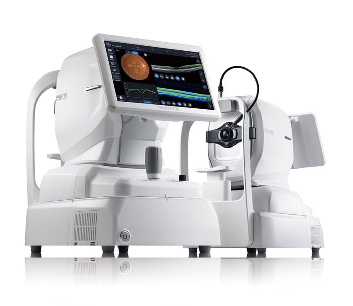
HOCT-1/1F with Angiography
All-in-One Optical Coherence Tomography with Fundus and Angiography
Features
-
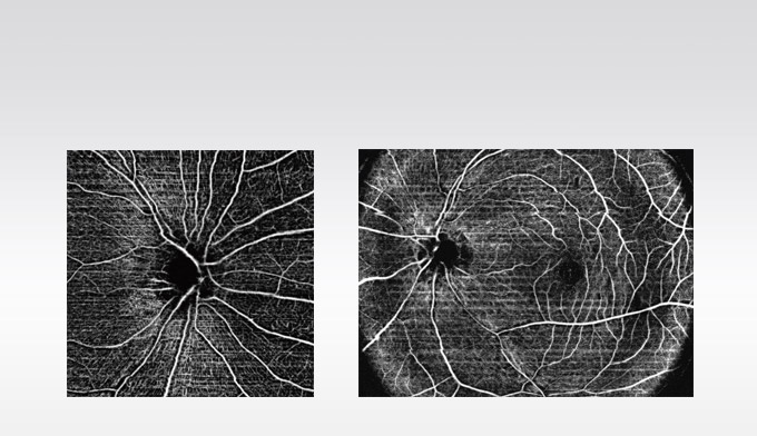
- Innovative Angiography
- Huvitz's own optical technologies, Real Time Tracking / Noise Cancelling / Motion Correction are inter-operating, and automatically analyzing & visualizing Retina, Microvessel of Choroid.
-

- High-Speed & High-Quality
- Provides High-speed Scan, High-quality Image by using Huvitz’s outstanding optical technology and innovative image software. Shows extensive information, such as 3D structure of Retina, Macula's thickness and separation, in a vivid image.
-
- One for All System
- By combining OCT-Angiography, Full Color Fundus Camera, and PC, it can generate high resolution images providing multi-purpose functions for diagnosis. It saves both time and space by performing frontal view(Enface) of eye diseases, Tomography, cross-compare and diagnosis in a single run.
-

- Web Browsing System
- Patient's test data can be analyzed anywhere on the Internet. You can check and analyze all data of HOCT through Web Browser such as Internet Explorer, Safari, Chrome without installing special software separately.
-

- Detailed Report
-
Provides patient's pathological structure and relevant & important data in easy-to-read format and also can print out the report on analysis screen.
Analysis results can be viewed via Web Browser and printed out with different types of reports.
-
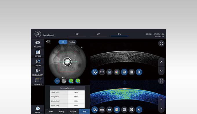
- Anterior Measurement
- Anterior Segment Module allows measurement and analysis of cornea thickness, angle and 3D image. It helps users work more efficiently by acquiring both anterior and posterior in one place.
-

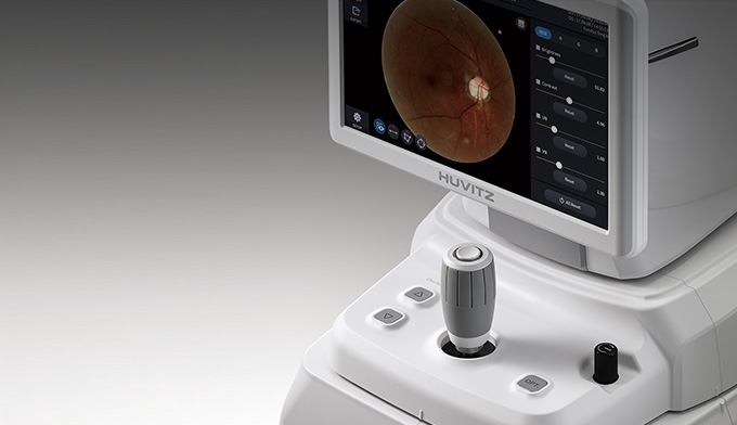
- Full Color Fundus Camera
- Color Retinal Images optimized with high-resolution and contrast are very useful in analysis and clinical diagnosis. Best images are provided by Low intensity of flash, fast capture speed, quiet operation, small pupil mode and automatic flicker detection.
Specifications
| Type | Spectral Domain OCT, Fundus Digital Photography | |
|---|---|---|
| OCT | Resolu tion in Tissue | 20um(Lateral), 6~7um(z-axis) at index 1.36 |
| Scan Speed | Max. 68,000 A-scan/sec. | |
| Scan Range | X: 6~12mm, Y: 6~9mm, Z: 2.34mm | |
| Acquisition Time of 3D Image | 1.4 sec.(Normal Mode, A512 x B96) | |
| Minimum Pupil Diameter | 2.5mm | |
| Light Source | 840nm | |
| Optical Power at Cornea | ≤650uW | |
| Scan Pattern | Macular: Macular Line, Macular Cross, Macular Radial, M a c u lar 3D, Macular Raster Disc: Disc Circle, Disc Radial, Disc 3D, Disc Raster |
|
| Angiography Range | X: 3~12mm, Y: 3~9mm(Optional) | |
| Angiography Map | Superficial, Deep, Outer, Choriocapillary, Retina, Custom, Enface, Thickness, Depth coded Map | |
| Angiography Analysis | FAZ, Vessel density Tools(Optional) | |
| Fundus | Camera | Built-in 12 Megapixel, Color |
| Angle of View | 45° | |
| Minimum Pupil Diameter | 4.0mm(Normal Mode), 3.3mm(Small Pupil Mode) | |
| Flash Light | White Light, 10 Levels | |
| Resolution |
Center: 60 Line pair/mm Middle: 40 Line pair/mm Periphery: Line pair/mm |
|
| Common | Working Distance | 33mm |
| Display | 12.1 inch, 1280x800 pixel, Touch Panel Color LCD | |
| Dioptic Compensation |
-33D~+33D Total -13D~+13D with No Compensation Lens +7D~+33D with Plus Compensation Lens -33D~-7D with Minus Compensation Lens |
|
| Internal Fixation Target | LCD(internal), White LED(external) | |
| Horizontal Movement | 70mm(back and forth), 100mm(left and right) | |
| Vertical Movement | 30mm | |
| Chinrest Movement | 62mm(up and down), Motorized | |
| Auto Tracking |
30mm(up and down) 10mm(right and left) 10mm(back and forth) |
|
| Network | DICOM File support (Need to be customized) | |
| PC | Built ni Computer | |
| Power Supply | AC100 -240V, 50/60Hz, 1.6-0.7A | |
| Dimensions / Mass | 330(W)x542(D)x521(H)mm / 30kg | |
| Accessories(Optional) | ||
| Anterior Segment Module |
Scan Pattern | ACA Line, Anterior Radial |
| Scan Range | 6~9mm(width), 2.3mm(depth) | |
| Software Analysis | Corneal Layers, Thickness Map, Thickness & Angle | |
| Wide Anterior Segment Module |
Scan Pattern | ACA Line, Anterior Radial |
| Scan Range | 16mm(width), 2.3mm(depth) | |
| Web-Viewer | Web-based, Multi users can be accesible. Progression Analysis, Comparison Analysis, 3D Analysis |
|
| Designs and details above can be changed without prior notice for the purposes of improvement. | ||