제품소개
-
- HIGH-SPEED & HIGH-QUALITY
- 빠른 Scan Speed, Smart Viewing Technology의 영상처리 알고리즘으로 안구의 움직임을 감지, 68,000A-scan/sec의 속도로 스캔하고 보정하여 매우 뛰어난 품질의 광학 이미지를 생성합니다.
-
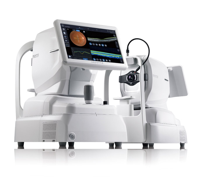
- One for All System & User Friendly
- OCT, Full Color Fundus Camera, PC가 결합한 일체형으로 고해상 이미지를 생성, 진단의 다목적 기능을 제공합니다. 또한 사용자의 측정 숙련도에 따른 영상 화질의 편차를 최소화해 신뢰도 높은 데이터를 획득합니다.
-
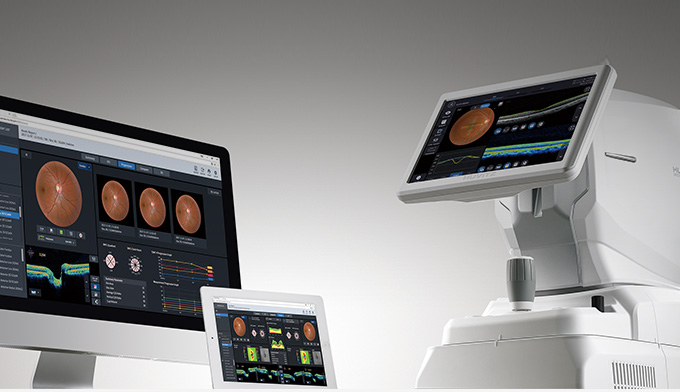
- Web Browsing System
- 환자의 검사 데이터는 인터넷을 사용하는 어느 곳에서라도 Internet Explorer, Safari, Chrome 등과 같은 Web Browser를 통해 HOCT의 모든 데이터를 분석할 수 있습니다.
-
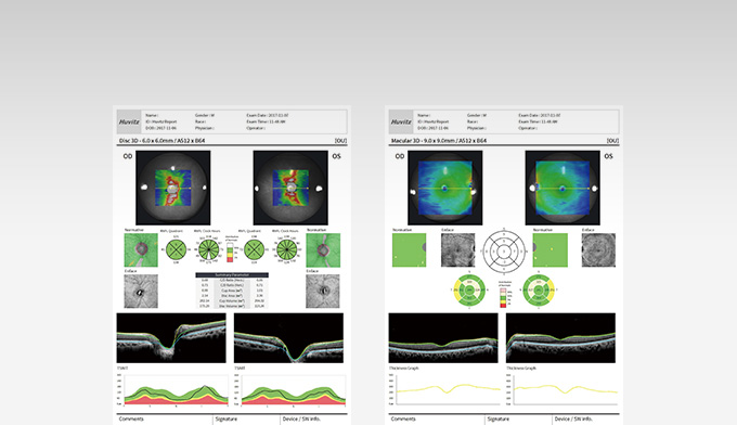
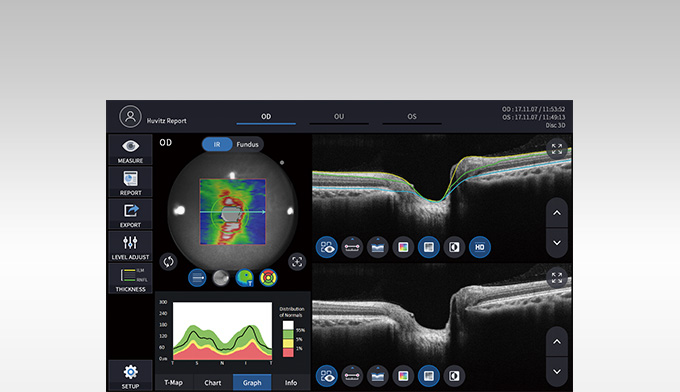
- DETAILED REPORT
- 병리적 구조 및 관련성 높은 중요 데이터를 읽기 쉬운 형식으로 제공하며 분석 화면에 설정된 형태로 인쇄가 가능합니다. 분석 결과는 Web Browser를 통해 볼 수 있고 여러 종류의 보고서로 인쇄할 수 있습니다.
-
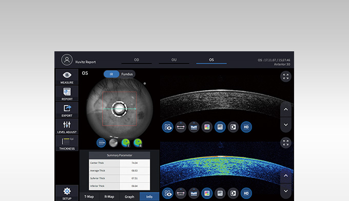
- ANTERIOR MEASUREMENT
- Anterior Segment Module를 장착하면 Cornea의 두께, ACA 각도, 3D Image를 측정, 분석할 수 있습니다. HOCT는 합리적인 올인원 OCT로서 한 자리에서 Anterior와 Posterior를 모두 획득하여 더욱 효율적입니다.
-
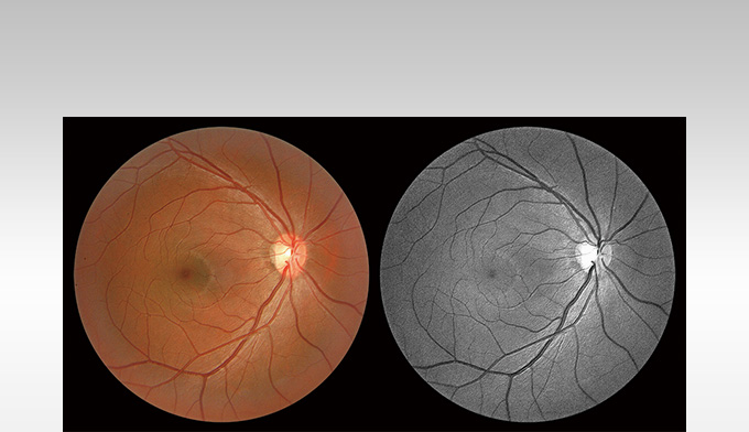
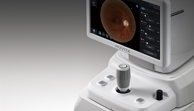
- FULL COLOR FUNDUS CAMERA
- High-Resolution, Contrast가 조율된 Color Retinal Images를 획득함으로써 분석과 임상 진단에 유용합니다. 낮은 플래시 강도, 빠른 캡처 속도, 조용한 작동, Small Pupil Mode 및 자동 깜박임 감지 등, 최상의 이미지를 캡처하기 위해 모두 담았습니다.
제품사양
| Type | SD-OCT / Fundus | ||
|---|---|---|---|
| OCT | Resolution(in Tissue) | Z :6~7um, XY:20um | |
| A scan Rate | 68,000 A-scan/sec. | ||
| Scan Range | Fundus] X:6-12mm, Y:6-9mm, Z:2.34mm [Cornea] X,Y:6-9mm |
||
| 3-D Acquisition Time | 1.4s(Fastest mode, A512 x B96) | ||
| Min. Pupil Diameter | Ø2.5mm | ||
| Light Source Wavelength | SLD 840nm | ||
| Optical Power at Cornea | ≤650uW | ||
| Scan Pattern | Macular Line, Macular Cross, Macular Radial, Macular Raster, Macular 3D, Disc Circle, Disc Radial, Disc Raster, Disc 3D | ||
| Fundus | Camera | Color, Resolution 12MP | |
| FOV | 45° | ||
| Min. Pupil Diameter | Normal:Ø4mm / Small Pupil:Ø3.3mm | ||
| Flash | LED | ||
| Resolution(on Fundus) | Center:60 lines/mm or more Middle(r/2):40 lines/mm or more Middle(r):25 lines/mm or more |
||
| Common | Working Distance | 33mm | |
| LCD Size | 12.1", Resolution 1280x800 | ||
| Dioptic Compensation | Full Range:-33 to +33D -33 to -7D with Minus Compensation Lens +7 to +33D with No Compensation Lens |
||
| Fundus Surface Imaging | NIR / Enface, FOV : 40°x30° | ||
| Internal Fixation Lamp | LCD | ||
| Horizontal Movement | 70mm (back and forth) / 100mm (left and right) | ||
| Vertical Movement | 30mm | ||
| Chinrest Movement | 62mm, Motorized | ||
| Auto Alignment | X,Y for Positioning, Z for Working Distance | ||
| Auto Focusing | Diopter Adjustment for Focusing | ||
| Network | DICOM File support (Need to be customized) | ||
| Built in Computer | O | ||
| Power Supply | AC100 ~ 240V, 50/60Hz, 1.6A~0.7A | ||
| Dimensions / Mass | 330(W)x542(D)x521(H)mm / 30kg | ||
| Optional Accessories | Anterior Segment Adapter, Web Viewer | ||
| Anterior Segment Module(optional) |
Scan Patterns | ACA Line, Cornea Radial, Cornea 3D | |
| Software Analysis | Corneal Layers, Thickness Map, Thickness & Angle | ||
| Web Viewer(optional) | Web-based, Multy users can be accesible Progression Analysis, Comparison Analysis, 3D Analysis |
||
| 상기 제품의 디자인과 세부 사항은 기능 향상을 위해 사전 예고없이 변경될 수 있습니다. | |||